
Cellular Adaptation in Pathology: Hypertrophy, Hyperplasia, Atrophy, Metaplasia and Atypia
Cells normally maintain a stable internal environment despite constant changes around them. However, when a cell is exposed to increased workload, hormonal stimulation, reduced blood supply, poor nutrition, chronic irritation, or other forms of stress, it may need to adjust its structure and function to survive. These changes are known as cellular adaptations.
Cellular adaptation is an important concept in general pathology because it helps explain how tissues respond to stress before permanent cell injury develops. Some adaptations are completely normal, while others occur because of disease or prolonged environmental stress.
The four major forms of cellular adaptation are hypertrophy, hyperplasia, atrophy, and metaplasia. Although atypia is often discussed in the same context, it is different from these adaptations. Atypia refers to abnormal cellular or nuclear features and is commonly associated with pathological processes such as dysplasia.
This article explains these changes with mechanisms, clinical examples, flowcharts, and important examination points for medical students and nursing students.
Cellular Adaptation in Pathology
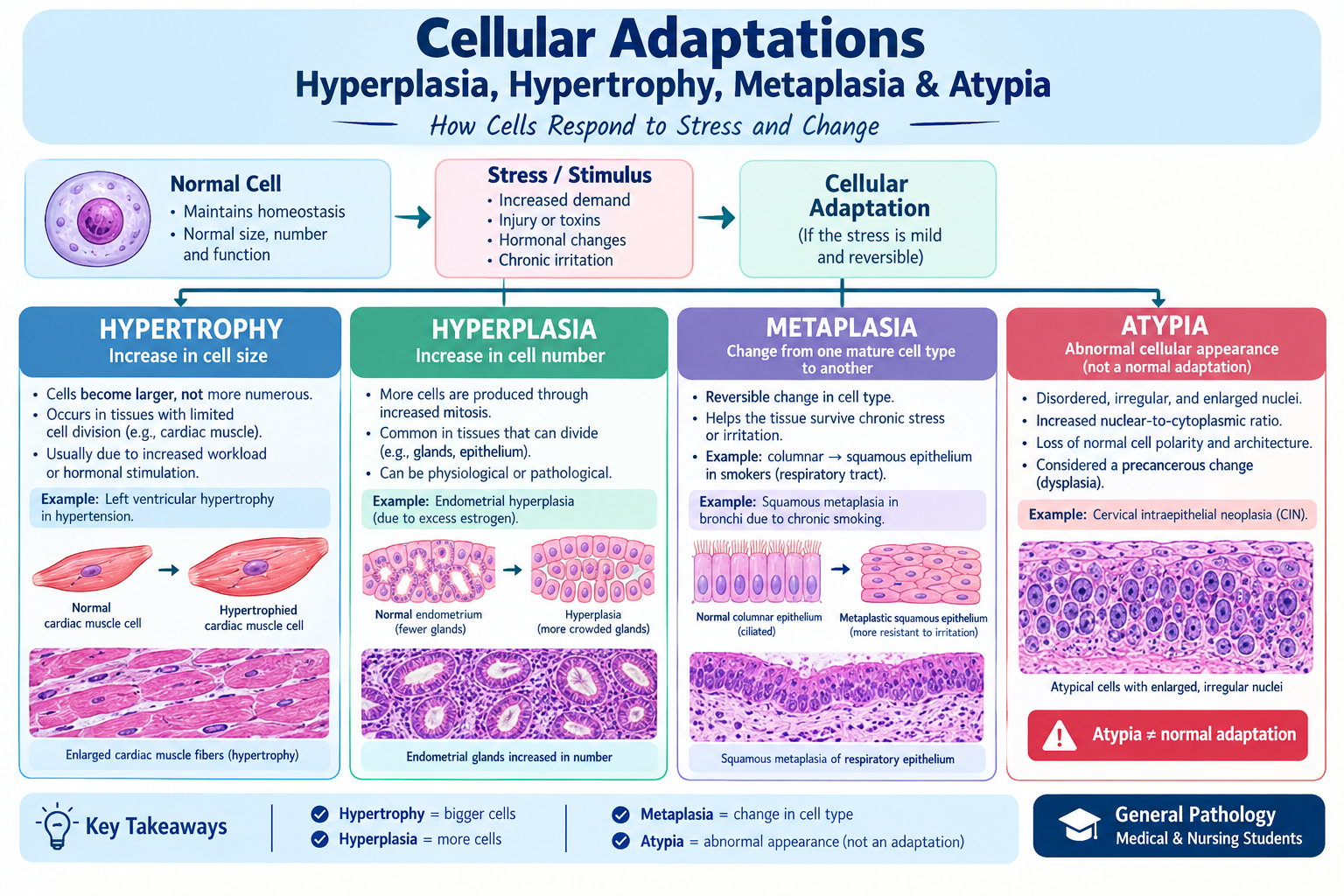
Educational medical illustration showing the major cellular adaptations discussed in general pathology, including hypertrophy, hyperplasia, atrophy, and metaplasia, with atypia presented separately as an abnormal cellular feature.
What Is Cellular Adaptation?
Cellular adaptation is a reversible change in the size, number, phenotype, metabolic activity, or function of cells in response to changes in their environment.
The purpose is simple: the cell attempts to cope with a new condition while remaining alive and functional.
For example, a person with long-standing hypertension has to pump blood against increased resistance. Cardiac muscle cells respond to this increased workload by becoming larger. This produces cardiac hypertrophy.
Similarly, when a tissue needs to produce more cells because of hormonal stimulation or increased demand, it may undergo hyperplasia.
These responses are useful because they allow cells to adjust rather than immediately becoming injured.
Cellular response to stress
Normal cell
↓
Change in environment or increased stress
↓
Cellular adaptation
↓
Cell survives and adjusts
If the stress becomes excessive or persists for too long:
Persistent/severe stress
↓
Cell injury
↓
Reversible injury or irreversible injury
↓
Cell death
The outcome depends on the severity and duration of the stress and on the ability of the particular tissue to adapt.
Major Types of Cellular Adaptation
The four classic cellular adaptations are:
- Hypertrophy – increase in cell size
- Hyperplasia – increase in cell number
- Atrophy – decrease in cell size
- Metaplasia – replacement of one mature cell type by another
Atypia is considered separately because it is an abnormal morphological finding rather than a classic adaptive response.
| Adaptation | Main change | Common example |
| Hypertrophy | Cells become larger | Left ventricular hypertrophy |
| Hyperplasia | Number of cells increases | Endometrial hyperplasia |
| Atrophy | Cells become smaller | Muscle wasting after immobilization |
| Metaplasia | One mature cell type is replaced by another | Respiratory squamous metaplasia |
1. Hypertrophy
Hypertrophy is an increase in the size of individual cells, which usually causes enlargement of the affected tissue or organ.
The important point is that hypertrophy involves larger cells rather than simply producing more cells.
It commonly develops when cells experience increased mechanical workload or hormonal stimulation. The cells respond by producing more structural and functional proteins, allowing them to handle the increased demand.
How hypertrophy develops
Increased workload or hormonal stimulation
↓
Mechanical and chemical signals activate intracellular pathways
↓
Increased protein synthesis
↓
Increase in cellular components
↓
Cell enlargement
↓
Hypertrophy
Physiological Hypertrophy
Not all hypertrophy is caused by disease. It can be a normal response to increased demand.
Skeletal muscle
Resistance exercise places repeated stress on skeletal muscle fibers. The cells respond by increasing their protein content and becoming larger.
This is why regular strength training can increase muscle mass.
Uterus during pregnancy
During pregnancy, hormonal stimulation contributes to enlargement of uterine smooth muscle cells. Increased cell proliferation may occur at the same time, so the enlargement of the uterus involves both hypertrophy and hyperplasia.
Pathological Hypertrophy
A classic example is left ventricular hypertrophy caused by chronic hypertension.
When blood pressure remains high, the left ventricle has to generate greater force to eject blood into the systemic circulation. Cardiac muscle cells respond by increasing their size.
Hypertension and cardiac hypertrophy
Chronic hypertension
↓
Increased pressure load on the left ventricle
↓
Increased workload of cardiac muscle
↓
Increased protein synthesis
↓
Enlargement of cardiac muscle cells
↓
Left ventricular hypertrophy
Initially, this response helps the heart cope with the increased workload. However, persistent hypertrophy can eventually become harmful. The enlarged myocardium has increased metabolic requirements and may develop functional abnormalities over time.
This illustrates an important principle of pathology: an adaptation that is initially beneficial can become detrimental when the underlying stress continues.
2. Hyperplasia
Hyperplasia is an increase in the number of cells in a tissue or organ caused by increased cell proliferation.
Unlike hypertrophy, the defining change is cell number.
Hyperplasia can occur in tissues containing cells that are capable of dividing. It may be triggered by hormones, growth factors, or compensatory signals.
Mechanism of hyperplasia
Growth stimulus
↓
Activation of growth-related signaling
↓
Cells enter the cell cycle
↓
Cell proliferation increases
↓
Cell number increases
↓
Hyperplasia
Physiological Hyperplasia
Physiological hyperplasia occurs during normal body processes.
Hormonal hyperplasia
Hormones can stimulate the proliferation of cells in tissues such as the breast and endometrium.
During pregnancy, for example, hormonal stimulation promotes proliferation of glandular epithelial cells in the breast, helping prepare the tissue for lactation.
Compensatory hyperplasia
The liver has a remarkable capacity for regeneration. Following partial loss of liver tissue, the remaining hepatocytes can proliferate in response to growth signals, helping restore the functional mass of the organ.
This is called compensatory growth.
Pathological Hyperplasia
Pathological hyperplasia occurs when growth-promoting signals become excessive or inappropriate.
One example is endometrial hyperplasia, which can occur with prolonged or excessive estrogenic stimulation.
Excessive estrogenic stimulation
↓
Increased endometrial cell proliferation
↓
Increased number of endometrial cells
↓
Endometrial hyperplasia
The clinical significance varies according to the type of hyperplasia. Some forms are associated with an increased risk of endometrial carcinoma and therefore require appropriate clinical assessment.
Does hyperplasia mean cancer?
No.
Hyperplasia is not the same as neoplasia. It is generally a regulated response to a stimulus. However, certain pathological forms of hyperplasia may be associated with an increased risk of malignant transformation.
Hypertrophy vs Hyperplasia
This distinction is particularly important in examinations.
Hypertrophy = bigger cells
Hyperplasia = more cells
An organ can sometimes enlarge through both processes.
For example, hormonal stimulation during pregnancy contributes to both hypertrophy and hyperplasia of uterine smooth muscle.
3. Atrophy
Atrophy is a reduction in the size of cells due to loss of cellular components. It commonly results in a smaller tissue or organ and reduced functional capacity.
An atrophic cell is not necessarily dead. Instead, it has adjusted to reduced demand or limited resources by decreasing its cellular components and metabolic activity.
Common causes include:
- Reduced workload
- Denervation
- Reduced blood supply
- Inadequate nutrition
- Loss of hormonal stimulation
- Aging
- Pressure
Atrophy from Reduced Workload
A familiar clinical example is skeletal muscle atrophy following prolonged immobilization.
When a limb remains inactive for an extended period, muscle fibers receive less stimulation and have a lower functional demand.
Immobilization
↓
Reduced muscle workload
↓
Reduced protein synthesis
Increased protein degradation
↓
Loss of cellular proteins and organelles
↓
Muscle fiber atrophy
This is one reason prolonged bed rest can result in significant muscle wasting.
Denervation Atrophy
Muscle tissue also depends on normal nerve stimulation.
When the nerve supplying a muscle is damaged, the muscle loses normal neural input and can progressively decrease in size.
Nerve damage
↓
Loss of normal stimulation
↓
Reduced muscle activity
↓
Increased protein degradation
↓
Loss of muscle mass
↓
Denervation atrophy
Atrophy from Poor Nutrition
Severe nutritional deficiency can cause the body to break down stored proteins and other cellular components to meet its energy requirements.
This contributes to muscle wasting and generalized tissue atrophy.
Atrophy from Reduced Blood Supply
Chronic reduction in blood supply can limit the delivery of oxygen and nutrients. Cells may respond by reducing their metabolic activity and size.
However, severe ischemia is different. When oxygen deprivation becomes sufficiently severe or prolonged, adaptation may no longer protect the cell and cell injury or cell death can occur.
Atrophy and Aging
Some degree of tissue atrophy occurs as part of aging.
Age-related changes can involve:
- Reduced protein synthesis
- Accumulation of cellular damage
- Altered metabolism
- Reduced regenerative capacity
- Changes in organ function
The extent and clinical significance vary among different tissues.
Mechanisms of Atrophy
Atrophy involves a balance between the production and breakdown of cellular components.
Two important mechanisms are:
Reduced protein synthesis
Cells produce fewer proteins needed to maintain their structure and function.
Increased protein degradation
Proteins are broken down more rapidly. The ubiquitin-proteasome system is particularly important in protein degradation, while autophagy contributes to the recycling of cellular components.
Atrophy flowchart
Reduced workload / denervation / malnutrition / reduced blood supply / hormonal withdrawal
↓
Reduced protein synthesis
Increased protein degradation
↓
Loss of cellular components
↓
Reduced cell size
↓
Atrophy
4. Metaplasia
Metaplasia is a reversible change in which one mature cell type is replaced by another mature cell type that is better able to tolerate a persistent environmental stress.
It is commonly associated with chronic irritation or inflammation.
The new cell type may be more resistant to the stressful environment, but it may not perform all of the specialized functions of the original tissue.
How Does Metaplasia Occur?
A common misconception is that one mature cell simply transforms directly into another mature cell.
Instead, metaplasia is generally associated with reprogramming of stem cells or undifferentiated progenitor cells within the tissue.
Signals from the local environment alter the pattern of differentiation.
Metaplasia flowchart
Chronic irritation or persistent stress
↓
Changes in local cellular signals
↓
Reprogramming of stem/progenitor cells
↓
Altered differentiation
↓
Development of a different mature cell type
↓
Metaplasia
Squamous Metaplasia in the Respiratory Tract
One of the classic examples is associated with chronic cigarette smoking.
Normal respiratory epithelium contains specialized ciliated columnar cells that help move mucus and trapped particles out of the airways.
Chronic exposure to cigarette smoke can cause persistent irritation.
The epithelium may respond by developing squamous metaplasia.
Cigarette smoke
↓
Chronic irritation
↓
Persistent epithelial stress
↓
Reprogramming of progenitor cells
↓
Change in epithelial differentiation
↓
Squamous metaplasia
Although squamous epithelium can tolerate certain forms of irritation better, the tissue loses some of the specialized functions of normal respiratory epithelium, including effective mucociliary clearance.
Why Is Metaplasia Important?
Metaplasia is generally reversible when the underlying stimulus is removed.
However, persistent irritation can produce additional pathological changes. In some tissues and disease settings, long-standing metaplasia can be associated with dysplasia and an increased risk of cancer.
This does not mean that every metaplastic change becomes malignant.
The progression is influenced by the cause, duration of exposure, tissue involved, and other genetic and environmental factors.
Metaplasia vs Dysplasia
These terms are often confused.
Metaplasia
One mature cell type is replaced by another mature cell type.
Dysplasia
There is disordered cellular growth and maturation, usually accompanied by cytological abnormalities and architectural disorganization.
Neoplasia
There is abnormal clonal cell proliferation associated with genetic alterations.
Therefore:
Metaplasia is not cancer, and metaplasia is not synonymous with dysplasia.
However, persistent pathological conditions can sometimes produce a sequence involving metaplasia, dysplasia, and eventually malignancy.
5. Understanding Atypia
Atypia refers to abnormal cellular or nuclear features that differ from those expected in normal tissue.
Unlike hypertrophy, hyperplasia, atrophy, and metaplasia, atypia is not one of the four classic cellular adaptations.
It is a descriptive pathological term used when cells appear abnormal under microscopic examination.
Features of Cellular Atypia
Depending on the condition, atypical cells may show:
Enlarged nuclei
The nucleus may be larger than expected.
Hyperchromasia
Nuclei may appear darker because of increased or altered chromatin.
Increased nuclear-to-cytoplasmic ratio
The nucleus occupies a greater proportion of the cell.
Pleomorphism
Cells and nuclei may vary in size and shape.
Abnormal mitotic figures
Some pathological lesions contain abnormal forms of cell division.
Loss of normal organization
Cells may become irregularly arranged within the tissue.
These findings are interpreted in combination rather than in isolation.
Atypia and Dysplasia
Atypia and dysplasia are closely related but should not be treated as identical terms.
Atypia mainly describes abnormal cellular morphology.
Dysplasia refers to disordered growth and maturation and often includes both cellular atypia and architectural abnormalities.
In epithelial tissues, dysplasia can be clinically important because certain grades and patterns may represent precancerous changes.
However, dysplasia is not automatically invasive cancer, and some dysplastic lesions can regress.
Cellular Adaptation vs Atypia
| Feature | Cellular adaptation | Atypia |
| Meaning | Response to environmental change or stress | Abnormal cellular morphology |
| Classic examples | Hypertrophy, hyperplasia, atrophy, metaplasia | Nuclear and cellular abnormalities |
| Normal physiological response? | Can be | No |
| Reversible? | Often | Depends on the cause |
| Associated with disease? | Sometimes | Commonly |
| Directly equal to cancer? | No | No |
When Does Adaptation Become Harmful?
An important concept in pathology is that adaptation is not always permanently protective.
A cell can successfully adapt to a stimulus for a period of time, but continued exposure may eventually cause damage.
Consider the following example:
Chronic hypertension
↓
Increased workload
↓
Cardiac hypertrophy
↓
Initial compensation
↓
Persistent pressure overload
↓
Structural and functional abnormalities
↓
Possible cardiac dysfunction
The same principle applies to chronic irritation and metaplasia.
Persistent irritant
↓
Metaplasia
↓
Continued exposure
↓
Additional cellular abnormalities
↓
Possible dysplasia in susceptible tissues
Thus, removing the underlying cause is often an important part of preventing progression.
Cellular Adaptation and Cell Injury
The relationship between adaptation and injury can be summarized as follows:
NORMAL CELL
↓
STRESS OR INCREASED DEMAND
↓
CAN THE CELL ADAPT?
↓
┌───────────────┐
│ YES │
└───────────────┘
↓
CELLULAR ADAPTATION
↓
┌────────┬──────────┬────────┬───────────┐
↓ ↓ ↓ ↓
Hypertrophy Hyperplasia Atrophy Metaplasia
↓
CELL SURVIVES
If the stress is too severe:
SEVERE OR PERSISTENT STRESS
↓
CELL INJURY
↓
┌───────┴────────┐
↓ ↓
Reversible Irreversible
injury injury
↓ ↓
Recovery Cell death
This framework helps connect the topic of cellular adaptation with the broader subject of cell injury and cell death.
Clinical Examples at a Glance
Hypertrophy
Condition: Chronic hypertension
Tissue: Left ventricular myocardium
Change: Cardiac muscle cells become larger
Hyperplasia
Condition: Excessive hormonal stimulation
Tissue: Endometrium
Change: Increased number of endometrial cells
Atrophy
Condition: Prolonged immobilization
Tissue: Skeletal muscle
Change: Muscle fibers become smaller
Metaplasia
Condition: Chronic cigarette smoke exposure
Tissue: Respiratory epithelium
Change: Ciliated columnar epithelium is replaced by squamous epithelium
Atypia
Finding: Abnormal cellular and nuclear morphology
Associated conditions: Dysplasia and neoplasia, among others
Important point: Atypia is not itself one of the classic cellular adaptations
High-Yield Points for Medical and Nursing Students
- Hypertrophy means an increase in cell size.
- Hyperplasia means an increase in cell number.
- Atrophy means a decrease in cell size and cellular components.
- Metaplasia means replacement of one mature cell type by another mature cell type.
- Hypertrophy commonly occurs in response to increased workload.
- Hyperplasia requires increased cell proliferation.
- Hypertrophy and hyperplasia can occur together.
- Atrophy can result from disuse, denervation, inadequate nutrition, reduced blood supply, hormonal withdrawal, and aging.
- Metaplasia is usually a response to chronic irritation or stress.
- Metaplasia is generally reversible if the underlying stimulus is removed.
- Smoking can cause squamous metaplasia of respiratory epithelium.
- Metaplasia is not synonymous with cancer.
- Atypia is not one of the four classic cellular adaptations.
- Dysplasia involves disordered growth and maturation and often includes cellular atypia.
- Persistent or severe stress can overwhelm cellular adaptation and result in cell injury.
Easy Memory Trick
Remember the four major adaptations as:
H-H-A-M
H — Hypertrophy
H — Hyperplasia
A — Atrophy
M — Metaplasia
Then remember what changes:
Hypertrophy → Size increases
Hyperplasia → Number increases
Atrophy → Size decreases
Metaplasia → Cell type changes
And:
Atypia → Abnormal appearance
Frequently Asked Questions
What is cellular adaptation in pathology?
Cellular adaptation is a reversible adjustment in cell size, number, phenotype, metabolism, or function that allows cells to cope with changes in their environment.
What are the four main types of cellular adaptation?
The four classic types are hypertrophy, hyperplasia, atrophy, and metaplasia.
What is the difference between hypertrophy and hyperplasia?
Hypertrophy involves an increase in cell size, whereas hyperplasia involves an increase in cell number.
What is an example of hypertrophy?
Left ventricular hypertrophy caused by chronic hypertension is a classic example of pathological hypertrophy.
What is an example of hyperplasia?
Endometrial hyperplasia caused by excessive hormonal stimulation is an example of pathological hyperplasia.
What causes muscle atrophy?
Muscle atrophy can result from reduced use, prolonged immobilization, denervation, inadequate nutrition, and other conditions that reduce normal muscle maintenance.
What is metaplasia?
Metaplasia is a reversible adaptive change in which one mature cell type is replaced by another mature cell type better suited to tolerate a persistent stress.
Is metaplasia cancer?
No. Metaplasia is an adaptive response. However, persistent pathological conditions associated with metaplasia can increase the risk of further abnormal changes in some tissues.
What is atypia?
Atypia describes abnormal cellular or nuclear morphology, such as nuclear enlargement, hyperchromasia, pleomorphism, or an increased nuclear-to-cytoplasmic ratio.
Is atypia a type of cellular adaptation?
No. The classic adaptations are hypertrophy, hyperplasia, atrophy, and metaplasia. Atypia is a pathological description of abnormal cellular appearance.
Conclusion
Cellular adaptation is the cell’s way of responding to changes in its environment while attempting to maintain survival and function. It is one of the basic concepts needed to understand general pathology.
When cells face increased workload, they may enlarge through hypertrophy. When appropriate growth signals stimulate proliferation, the number of cells may increase through hyperplasia. Reduced workload or limited nutritional and hormonal support can lead to atrophy, while chronic irritation may result in metaplasia, in which one mature cell type is replaced by another.
Atypia should be considered separately. It describes abnormal cellular or nuclear morphology and may be seen in dysplastic or neoplastic processes.
The central principle is straightforward:
Cellular stress → adaptation → survival
But when the stress is too severe or continues for too long:
Persistent stress → cell injury → irreversible damage → cell death
Understanding this progression provides an important foundation for studying general pathology, cell injury, inflammation, dysplasia, cancer, and disease mechanisms.
References
Kumar, V., Abbas, A. K., & Aster, J. C. Robbins Basic Pathology (10th ed.). Elsevier.
Scott, J. Oxford Handbook of Clinical Pathology (2nd ed.). Oxford University Press.
This article is an educational summary based on standard general pathology concepts and is intended for medical and nursing education.