
1. Learning Objectives
After studying this topic, you should be able to:
- Define cellular adaptation, cell injury, and cell death.
- Explain how cells respond to physiological and pathological stress.
- Differentiate reversible and irreversible cell injury.
- Describe the major causes of cell injury.
- Explain the major biochemical mechanisms of cell injury.
- Describe the effects of hypoxia, ischemia, toxins, free radicals, and DNA damage.
- Differentiate necrosis and apoptosis.
- Explain the four major cellular adaptations:
- Hypertrophy
- Hyperplasia
- Atrophy
- Metaplasia
- Recognize important clinical examples of cellular injury.
2. Normal Cellular Homeostasis
A normal cell continuously adjusts its structure and function to maintain a relatively stable internal environment called homeostasis.
Cells must maintain:
- ATP production
- Normal ion concentrations
- Appropriate intracellular pH
- Protein synthesis
- DNA integrity
- Membrane integrity
- Normal mitochondrial function
- Removal of damaged cellular components
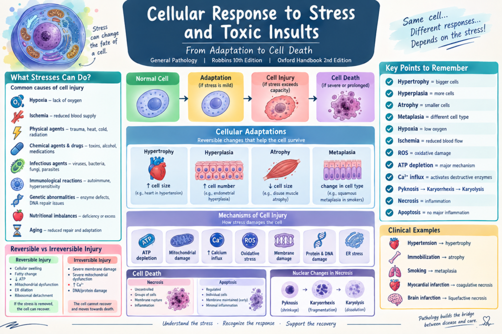
When cells encounter stress, they have several possible responses:
Normal cell → Adaptation → Reversible injury → Irreversible injury → Cell death
The outcome depends mainly on:
- Type of stress
- Severity
- Duration
- Type of cell affected
- Cell’s ability to adapt
Robbins emphasizes that mild or short-lived injury may be reversible, whereas severe, persistent, or rapidly developing injury may become irreversible and result in cell death.
3. Cellular Responses to Stress
There are three major outcomes:
A. Adaptation
The cell modifies its structure or function to survive the stress.
Examples:
- Hypertrophy
- Hyperplasia
- Atrophy
- Metaplasia
B. Cell Injury
Occurs when the stress exceeds the cell’s ability to adapt.
It may be:
Reversible injury
or
Irreversible injury
C. Cell Death
Severe or persistent injury can result in:
- Necrosis
- Apoptosis
Simplified flowchart
Stress
↓
Can the cell adapt?
→ Yes → Cellular adaptation
→ No → Cell injury
↓
Reversible?
→ Yes → Recovery
→ No → Cell death
4. Causes of Cell Injury
Important causes include:
1. Hypoxia
Hypoxia = inadequate oxygen availability to cells.
Causes include:
- Ischemia
- Cardiorespiratory failure
- Severe anemia
- Carbon monoxide poisoning
- Reduced oxygen-carrying capacity
2. Ischemia
Ischemia = reduced blood supply to a tissue.
It is particularly damaging because it causes:
- Reduced oxygen
- Reduced nutrients
- Reduced removal of metabolic waste
Therefore, ischemia can be more damaging than hypoxia alone.
3. Physical Agents
Examples:
- Trauma
- Heat
- Cold
- Radiation
- Electric shock
- Changes in atmospheric pressure
4. Chemical Agents and Drugs
Examples:
- Poisons
- Alcohol
- Certain medications
- Industrial chemicals
- Environmental pollutants
- Heavy metals
5. Infectious Agents
Examples:
- Viruses
- Bacteria
- Fungi
- Parasites
6. Immunological Reactions
Examples:
- Autoimmune diseases
- Hypersensitivity reactions
The immune response can itself damage tissues.
7. Genetic Abnormalities
Genetic defects can result in:
- Abnormal proteins
- Enzyme deficiencies
- Defective DNA repair
- Abnormal cellular metabolism
8. Nutritional Imbalances
Both deficiency and excess can cause cellular injury.
Examples:
- Protein-calorie malnutrition
- Vitamin deficiency
- Obesity
- Excessive lipid accumulation
9. Aging
Aging reduces the ability of cells to respond appropriately to stress and repair damage.
5. Cellular Adaptation
Cellular adaptation is a reversible change in cell size, number, phenotype, metabolic activity, or function that allows cells to survive altered environmental conditions.
The four major adaptations are:
| Adaptation | Main change | Example |
| Hypertrophy | ↑ Cell size | Cardiac muscle in hypertension |
| Hyperplasia | ↑ Cell number | Endometrial proliferation |
| Atrophy | ↓ Cell size | Disuse muscle atrophy |
| Metaplasia | Change in cell type | Respiratory epithelium in smokers |
These adaptations are important because they allow cells to maintain viability despite increased or altered demands.
6. Hypertrophy
Definition
Hypertrophy is an increase in the size of individual cells, resulting in enlargement of the affected organ or tissue.
The number of cells does not increase.
Common examples
Physiological hypertrophy
- Skeletal muscle enlargement during exercise
- Uterine smooth muscle enlargement during pregnancy
Pathological hypertrophy
- Left ventricular hypertrophy in hypertension
- Cardiac hypertrophy caused by valvular disease
Mechanism
Increased workload → mechanical/chemical signals → increased protein synthesis → increased cell size
Important point
Hypertrophy is particularly important in tissues where cells have limited ability to divide, such as cardiac muscle cells.
7. Hyperplasia
Definition
Hyperplasia is an increase in the number of cells in a tissue or organ.
It occurs in cells capable of division.
Types
Physiological hyperplasia
Hormonal
Example:
- Breast glandular tissue during pregnancy
Compensatory
Example:
- Liver regeneration after partial removal
Pathological hyperplasia
Usually caused by excessive hormonal or growth-factor stimulation.
Examples:
- Endometrial hyperplasia
- Benign prostatic hyperplasia
Important distinction
Hypertrophy = bigger cells
Hyperplasia = more cells
Both may occur together.
8. Atrophy
Definition
Atrophy is a decrease in the size of cells and often a reduction in the size of the affected organ.
Causes include:
- Reduced workload
- Loss of nerve supply
- Reduced blood supply
- Inadequate nutrition
- Loss of endocrine stimulation
- Aging
- Pressure
Example
A patient immobilized in bed for several weeks may develop skeletal muscle atrophy.
Mechanisms
Atrophy involves:
- Reduced protein synthesis
- Increased protein degradation
- Autophagy
9. Metaplasia
Definition
Metaplasia is a reversible change in which one mature cell type is replaced by another mature cell type better able to tolerate a persistent stress.
Example: Smoking
Normal respiratory epithelium:
Ciliated columnar epithelium
↓
Chronic irritation from cigarette smoke
↓
Squamous metaplasia
The new squamous epithelium is more resistant to irritation but loses some specialized functions, such as effective mucociliary clearance.
Important
Metaplasia is not cancer.
However, persistent pathological stimulation may increase the risk of further abnormal changes and, in some settings, malignancy.
10. Reversible Cell Injury
Reversible injury occurs when the damaging stimulus is removed before severe cellular damage develops.
The cell can return toward normal.
Major features
- Cellular swelling
- Fatty change
- Reduced ATP
- Mitochondrial dysfunction
- Endoplasmic reticulum dilation
- Ribosomal detachment
- Membrane changes
Mechanism
Injury
↓
Reduced ATP production
↓
Failure of Na⁺/K⁺ ATPase pump
↓
Na⁺ accumulates inside cell
↓
Water enters cell
↓
Cell swelling
Clinical/pathological terms
This may be called:
- Hydropic change
- Cellular swelling
12. Fatty Change
Fatty change = abnormal accumulation of triglycerides within cells.
It is particularly common in:
- Liver
- Heart
- Kidney
Important causes
- Alcohol
- Obesity/metabolic disorders
- Toxins
- Diabetes/metabolic disturbances
- Hypoxia
Example
Fatty liver may occur with chronic alcohol consumption or metabolic dysfunction.
13. Irreversible Cell Injury
If injury becomes sufficiently severe, the cell reaches a point of no return.
Major features include:
1. Severe mitochondrial dysfunction
The mitochondria can no longer produce adequate ATP.
2. Severe membrane damage
Cellular contents leak out.
3. Severe calcium influx
Intracellular Ca²⁺ rises and activates destructive enzymes.
4. Severe DNA/protein damage
This can activate pathways leading to cell death.
At this stage, the cell cannot recover.
14. Major Mechanisms of Cell Injury
Robbins describes several interconnected mechanisms.
The major mechanisms are:
- ATP depletion
- Mitochondrial damage
- Increased intracellular calcium
- Oxidative stress
- Membrane damage
- Protein and DNA damage
- Endoplasmic reticulum stress
15. ATP Depletion
ATP is essential for cellular survival.
ATP is required for:
- Ion pumps
- Protein synthesis
- Lipid synthesis
- Cellular transport
- Maintenance of membrane potential
ATP depletion causes:
↓ Na⁺/K⁺ pump activity
↓
Na⁺ enters cell
↓
Water follows Na⁺
↓
Cell swelling
ATP depletion also causes:
↓ Protein synthesis
↓
Ribosomal detachment from rough ER
↓
Reduced protein production
16. Mitochondrial Damage
Mitochondria are essential for ATP production.
Mitochondrial injury causes:
- Reduced ATP
- Increased reactive oxygen species
- Release of proteins that can activate apoptosis
Severe mitochondrial damage can therefore contribute to both necrosis and apoptosis.
17. Calcium Influx
Normally, cytoplasmic calcium concentration is kept very low.
Cell injury causes increased intracellular Ca²⁺.
Increased Ca²⁺ activates enzymes such as:
- Phospholipases
- Proteases
- Endonucleases
- ATPases
These enzymes damage:
- Cell membranes
- Cytoskeletal proteins
- DNA
- ATP stores
18. Oxidative Stress
Definition
Oxidative stress occurs when production of reactive oxygen species (ROS) exceeds the cell’s antioxidant defenses.
Important ROS include:
- Superoxide
- Hydrogen peroxide
- Hydroxyl radical
ROS can damage:
Lipids → membrane damage
Proteins → loss of protein function
DNA → mutations and cell death
19. Antioxidant Defenses
Cells have protective mechanisms against ROS.
Important antioxidants include:
- Vitamin E
- Vitamin C
- Glutathione
- Superoxide dismutase
- Catalase
Important concept
ROS production > antioxidant capacity → oxidative stress → cellular injury
20. Membrane Damage
Cell membranes are essential for:
- Maintaining ionic gradients
- Controlling cellular contents
- ATP production
- Communication
Damage to membranes can result in leakage of intracellular components.
Plasma membrane damage
→ Loss of cellular contents
→ Influx of ions and water
→ Cell swelling
→ Necrosis
Mitochondrial membrane damage
→ Loss of membrane potential
→ Reduced ATP
→ Cell death
21. Endoplasmic Reticulum Stress
The endoplasmic reticulum is involved in protein synthesis and folding.
Accumulation of misfolded proteins produces ER stress.
Cells respond through the unfolded protein response (UPR).
The UPR attempts to:
- Reduce new protein synthesis
- Increase protein folding capacity
- Remove abnormal proteins
If ER stress is severe or prolonged, it may contribute to apoptosis.
22. DNA Damage
DNA can be damaged by:
- Radiation
- ROS
- Chemicals
- Mutations
- Certain infections
Cells attempt to repair DNA.
If damage is too severe to repair, the cell may undergo apoptosis.
This is an important protective mechanism because it prevents severely damaged cells from surviving and transmitting mutations.
23. Toxic Cellular Injury
Toxic substances can injure cells by several mechanisms.
Direct toxicity
The toxic substance itself damages an important cellular component.
Examples include substances that directly damage:
- Cell membranes
- Mitochondria
- Proteins
- DNA
Indirect toxicity
Some chemicals are metabolized into highly reactive toxic compounds.
These metabolites may produce:
- ROS
- Membrane damage
- Protein damage
- DNA damage
Therefore:
Toxin → cellular metabolism → toxic metabolite → cellular damage
Robbins’ treatment specifically includes cell injury caused by toxins as one of the mechanisms discussed in this chapter.
24. Necrosis
Definition
Necrosis is uncontrolled cell death caused by severe cellular injury and is usually associated with inflammation.
Typical sequence:
Severe injury
↓
Loss of ATP and membrane integrity
↓
Cell swelling
↓
Membrane rupture
↓
Leakage of cellular contents
↓
Inflammatory response
25. Nuclear Changes in Necrosis
There are three classic nuclear changes:
Pyknosis
Nucleus becomes:
- Small
- Dark
- Condensed
Karyorrhexis
The condensed nucleus breaks into fragments.
Karyolysis
The nucleus fades and dissolves due to enzymatic degradation of DNA.
Sequence
Pyknosis → Karyorrhexis → Karyolysis
This is an important exam point.
26. Types of Necrosis
| Type | Typical example |
| Coagulative | Myocardial infarction |
| Liquefactive | Brain infarction, abscess |
| Caseous | Tuberculosis |
| Fat | Acute pancreatitis |
| Fibrinoid | Immune-mediated vascular injury |
| Gangrenous | Severe ischemic limb injury |
27. Apoptosis
Definition
Apoptosis is a regulated form of cell death in which individual cells are eliminated without the extensive inflammation typically associated with necrosis.
It is important in:
- Normal development
- Removal of unwanted cells
- Elimination of damaged cells
- Maintaining tissue homeostasis
Examples
Physiological:
- Removal of cells during embryonic development
- Elimination of excess cells
- Endometrial cell loss during menstruation
Pathological:
- DNA-damaged cells
- Cells infected by certain viruses
- Cells with misfolded proteins
28. Apoptosis vs Necrosis
| Feature | Apoptosis | Necrosis |
| Nature | Regulated | Usually uncontrolled |
| Number of cells | Individual cells | Often groups of cells |
| Cell size | Shrinks | Swells |
| Membrane | Generally maintained initially | Disrupted |
| Nuclear changes | Fragmentation | Pyknosis → karyorrhexis → karyolysis |
| Inflammation | Usually minimal/absent | Usually prominent |
| Physiological role | Very important | Mainly pathological |
29. Intracellular Accumulations
Cells can accumulate abnormal substances because of:
- Abnormal metabolism
- Defective transport
- Inadequate degradation
- Excessive intake
Examples include:
Lipids
Fatty change in liver.
Proteins
Protein accumulation in certain renal and other cellular disorders.
Glycogen
May accumulate in certain metabolic diseases.
Pigments
Examples:
- Lipofuscin
- Melanin
- Hemosiderin
- Bilirubin
30. Autophagy
Definition
Autophagy is a cellular process in which damaged or unnecessary cellular components are delivered to lysosomes for degradation and recycling.
It becomes particularly important during:
- Nutrient deprivation
- Cellular stress
- Starvation
Purpose
Autophagy allows the cell to recycle:
- Amino acids
- Fatty acids
- Other cellular components
It can therefore help cells survive periods of stress.
31. Cellular Aging
With aging, cells gradually accumulate:
- DNA damage
- Oxidative damage
- Abnormal proteins
- Mitochondrial dysfunction
Cells also have reduced capacity for:
- Repair
- Replication
- Adaptation
This contributes to cellular senescence and age-related tissue dysfunction.
32. High-Yield Clinical Examples
Example 1: Hypertension
Chronic hypertension
↓
Increased workload on heart
↓
Cardiac muscle adapts
↓
Hypertrophy
Example 2: Immobilization
Limb immobilized for several weeks
↓
Reduced workload
↓
Reduced protein synthesis + increased protein degradation
↓
Muscle atrophy
Example 3: Smoking
Chronic cigarette smoke exposure
↓
Persistent irritation
↓
Respiratory epithelial adaptation
↓
Squamous metaplasia
Example 4: Myocardial infarction
Coronary artery obstruction
↓
Ischemia
↓
Hypoxia + ATP depletion
↓
Severe cellular injury
↓
Coagulative necrosis
Example 5: Brain infarction
Cerebral ischemia
↓
Neuronal injury
↓
Liquefactive necrosis
33. Nursing/Medical Exam Quick Review
Remember: H-H-A-M
H = Hypertrophy → increased cell size
H = Hyperplasia → increased cell number
A = Atrophy → decreased cell size
M = Metaplasia → altered cell type
Reversible injury
Think:
SWELLING + FAT
- Cellular swelling
- Fatty change
Irreversible injury
Think:
MEMBRANE + MITOCHONDRIA
- Severe membrane damage
- Severe mitochondrial dysfunction
Necrosis
Think:
CELL SWELLS → MEMBRANE RUPTURES → INFLAMMATION
Apoptosis
Think:
CELL SHRINKS → FRAGMENTS → NO MAJOR INFLAMMATION
34. Most Important Differences for Exams
| Concept | Remember |
| Hypertrophy | Increase in cell size |
| Hyperplasia | Increase in cell number |
| Atrophy | Decrease in cell size |
| Metaplasia | One mature cell type changes to another |
| Reversible injury | Cell can recover |
| Irreversible injury | Cell cannot recover |
| Necrosis | Cell death with membrane disruption and inflammation |
| Apoptosis | Regulated cell death, usually without significant inflammation |
| Hypoxia | Reduced oxygen |
| Ischemia | Reduced blood supply |
| ROS | Cause oxidative damage |
| ATP depletion | Major mechanism of cellular injury |
| Ca²⁺ influx | Activates destructive enzymes |
| Pyknosis | Nuclear shrinkage |
| Karyorrhexis | Nuclear fragmentation |
| Karyolysis | Nuclear dissolution |
Core concept to remember
Normal cell → Stress → Adaptation
If stress becomes too severe:
Stress → Cell injury → Reversible injury → Irreversible injury → Cell death
The central principle from Robbins is that the type, severity, and duration of stress, together with the cell’s type and ability to adapt, determine whether the cell adapts, suffers reversible injury, or progresses to cell death.
[…] Cellular Adaptation in Pathology: Hypertrophy, Hyperplasia, Atrophy, Metaplasia and Atypia […]